
Product · Workflows
Built around the questions chemists actually ask.
14 ready-made workflows in 3 problem families. Each one is a tested chain of tools with its file formats and hand-offs already connected: add your inputs, check the estimate, and run.
Family 01 · 5 stages
Virtual screening
Find the compounds in a library most likely to bind your target, then rescore the best hits with a stronger method.
Screen a compound library against a target
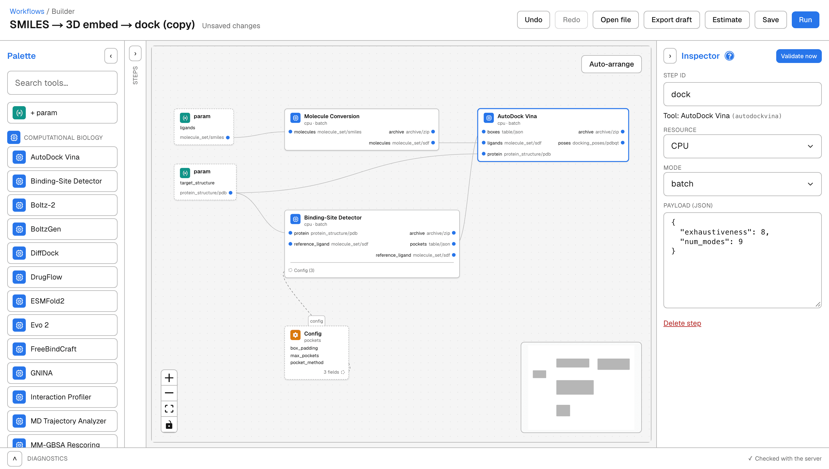
SMILES → 3D embed → dock
Convert SMILES to 3D structures, detect pockets, then dock with AutoDock Vina.
- You provide
- Molecules (SMILES), Protein structure (PDB)
- Runs on
- CPU
- Cost cap
- $10.00 default
Your library
Imatinib Nilotinib Dasatinib Your target
A pose in the pocket
- Target
- ABL1 kinase domain
- Structure
- PDB 2HYY · 2.4 Å
- Polar contacts
- 6
Family 02 · 6 stages
Structure prediction to docking
Dock against a protein with no solved structure: predict it from sequence, find its pockets, then dock.
Dock into a predicted structure without choosing a box
Sequence → fold → DiffDock
Fold a protein sequence with ESMFold2, dock a SMILES library blind with DiffDock, and keep the top-confidence poses.
- You provide
- Molecules (SMILES), Protein sequence
- Runs on
- A10 GPU · T4 GPU
- Cost cap
- $110.00 default
Your sequence
Its structure
A pose in the pocket
- Sequence
- 273 residues
- Structure
- PDB 2HYY · 2.4 Å
- Ligand
- Imatinib
Family 03 · 6 stages
Library design
Generate or enumerate new compounds for a target or an SAR series, then dock them.
Dock analogs enumerated from your SAR data
Free Wilson → 3D embed → dock
Enumerate analogs with Free Wilson, embed to 3D, detect pockets, then dock with AutoDock Vina.
- You provide
- Molecules (SMILES), Protein structure (PDB)
- Runs on
- CPU
- Cost cap
- $15.00 default
Your series
Imatinib Nilotinib Radotinib A combination not in it
Radotinib head · Imatinib tail A pose in the pocket
- Series
- 3 ABL inhibitors
- Shared core
- Phenylaminopyrimidine
- New combination
- C28H30N8O
Under the hood
Change any step, or build your own.
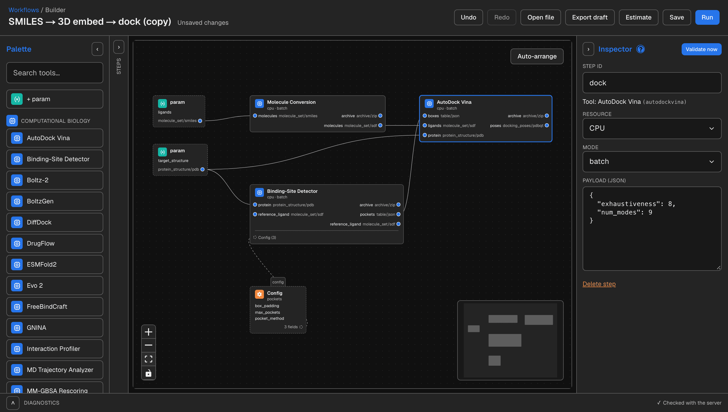
Every template opens in the visual builder. The same building blocks are available for your own workflows, and the builder checks each connection as you make it.
Bindings
Each step input is bound to one source:
| Binding | Connects to |
|---|---|
$param | A workflow input you fill in at launch: a file, a list, or a setting. |
$from | Another step's output port, e.g. the poses from a docking step. |
$artifact | A file already in your library, such as a result from an earlier run. |
$const | A fixed value written into the workflow. |
Typed ports
Every port carries a data kind, so a step only accepts what it can use. Known format conversions, such as SMILES to 3D SDF or CIF to PDB, run between steps automatically.
- Archive
- Docked poses
- Force-field system
- Kinetic mechanism
- Molecules
- Protein sequences
- Protein structure
- Sequences
- Structures
- Table
- Trained model